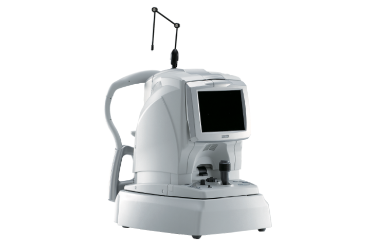
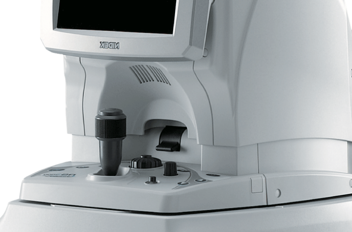
Optical Coherence Tomography
RS-3000 Advance II
Brand: Nidek – Japan
Providing a comprehensive solution for retina and glaucoma
- Retina Analysis
- Glaucoma Analysis
- AngioScan
- SLO
Brand: Nidek – Japan
Providing a comprehensive solution for retina and glaucoma
1. Retina Analysis
Selection of the appropriate OCT sensitivity allows acquisition of B-scan images through media opacities.
The tracing HD plus function traces involuntary eye movements to maintain the same scan location on the SLO image for accurate image capture. This function allows accurate averaging of up to 120 images. The tracing HD plus function combined with ultra fine sensitivity image capture results in high resolution and high contrast images of chorioretinal pathology.
Enhanced image function allows greater resolutions of vitreous retina images by adjusting brightness of weak OCT signals.
The select and rescan mode allows capture of an entire image of the retina with the macula map scan pattern and select a cross-sectional OCT image with the location of lesion from up to 256 images based on user preference.
Cross-sectional OCT images can be reacquired on the selected region with the tracing HD plus
function.
This mode is useful in efficiently obtaining a high-quality image of a region of interest.
2. Glaucoma Analysis
Wide area 9 x 9 mm normative database allows analysis of [NFL+GCL+IPL] thinning from optic disc to macula in a single report.
Anterior Chamber Angle
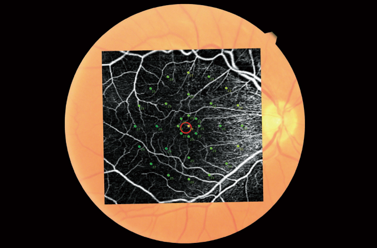
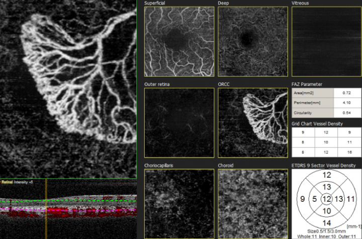
3. AngioScan
This non-invasive method does not require contrast dye injection for examination of the layer-by-layer microvasculature within the retina and choroid. Radial peripapillary capillary plexus (RPCP), superficial capillary plexus (SCP), internal capillary plexus (ICP) and deep capillary plexus (DCP) can be analyzed.
Images of the superficial capillary, deep capillary, outer retina and choroid can be displayed for clinical evaluation.
Tracing HD Plus
Selectable Definition
Two-, four- or eight-scan per line (2 HD, 4 HD, 8 HD) can be selected.
8 HD provides
Fine Mode
Fine mode OCT angiography results in high-resolution images to enhance diagnosis.
VIETCAN SERVICE & TRADING J.S.C
Address: Room 4A, JVPE Building, Quang Trung Software Park, Tan Chanh Hiep Ward, District 12, Ho Chi Minh City, Vietnam
License No. 0302438819, 11th additional issuance on March 27, 2018.
Place of issue: Department of Planning and Investment HCMC.
Phone: +84 28 6290 8200 – Email: [email protected]

Copyright by VietCan 2019