Retina Scan Duo™
Next Gen dual mode retinal imaging.

Next Gen dual mode retinal imaging.
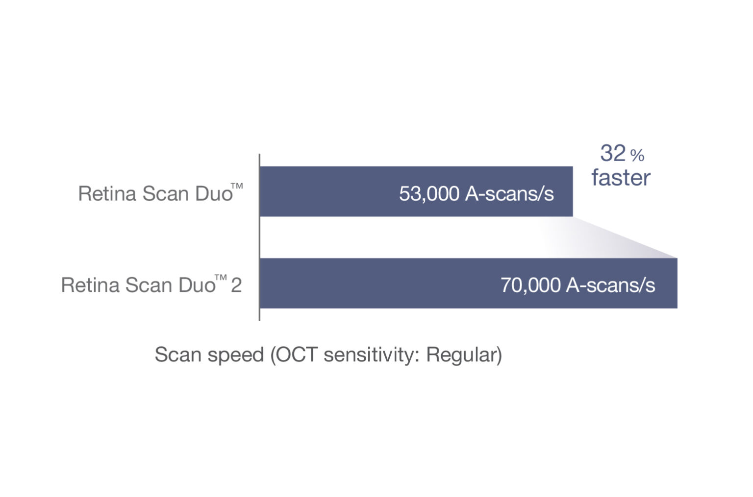
OCT
OCT images are captured at scan speeds of 70,000 A-scans/s which is 32% faster than acquisition with the Retina Scan Duo™ using Regular OCT sensitivity* .
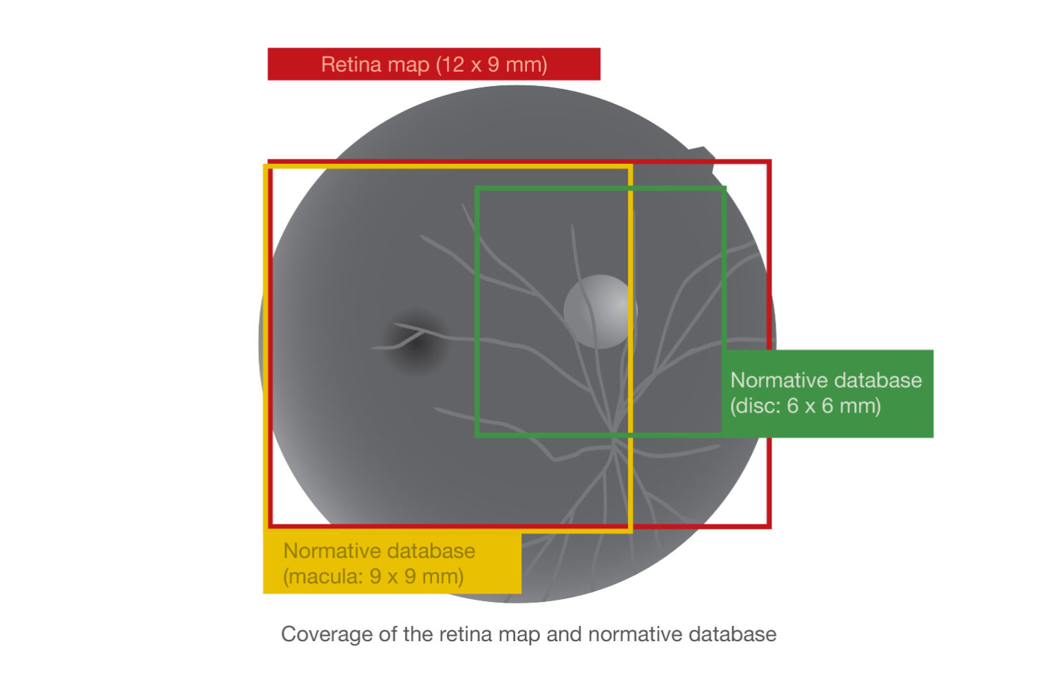
A 12 x 9 mm wide area image can be acquired. The retina map captures both the macula and disc in a single shot. The normative database provides a wide area color-coded map comparing the patient’s macular thickness to a population of normal eyes.
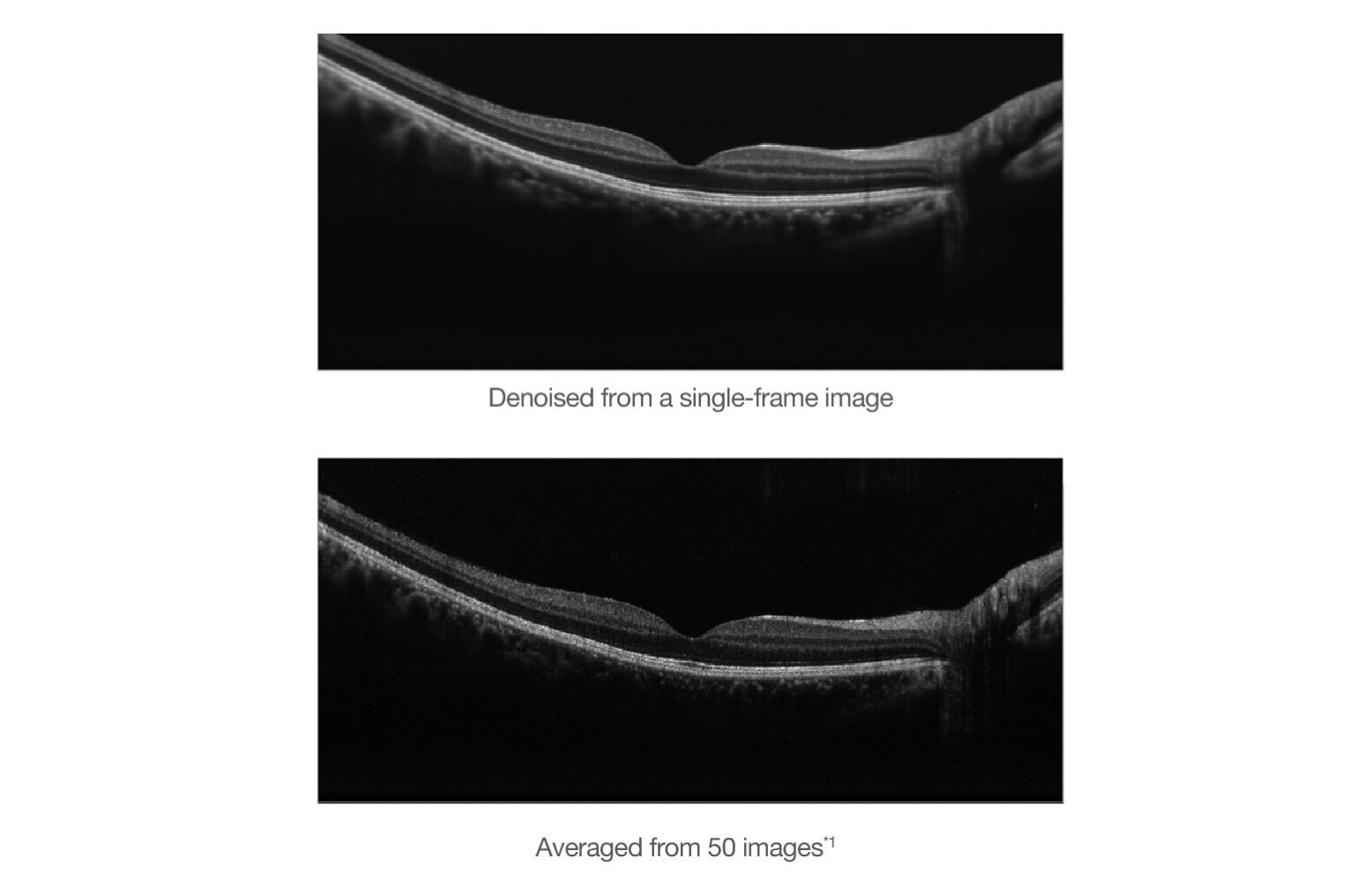
Denoising using deep learning
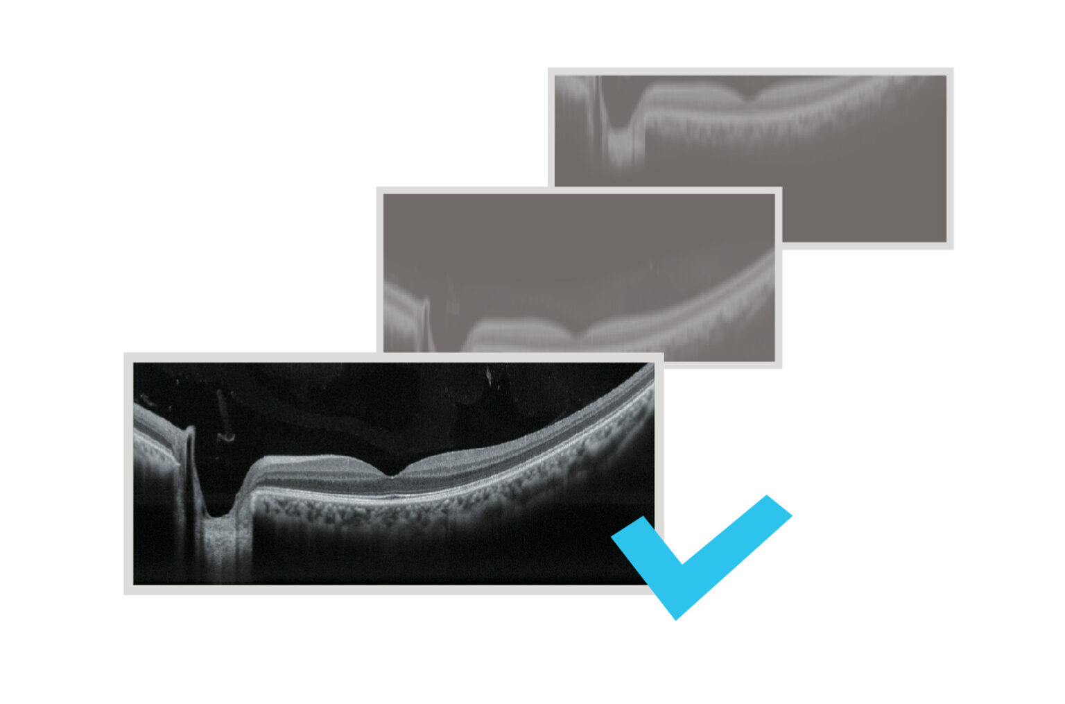
A new image enhancement technique using deep learning automatically displays a denoised image once B-scan acquisition is complete. With deep learning of a large data set of images averaged from 120 images, this denoising technique provides high definition images comparable to a multiple-image-averaging technique. The denoising function generates high definition images from a single frame while decreasing image acquisition time and increasing patient comfort.
Fundus Camera
The Retina Scan Duo™ 2 includes a built-in 12-megapixel CCD camera, producing high quality fundus images with a 45° angle of view.
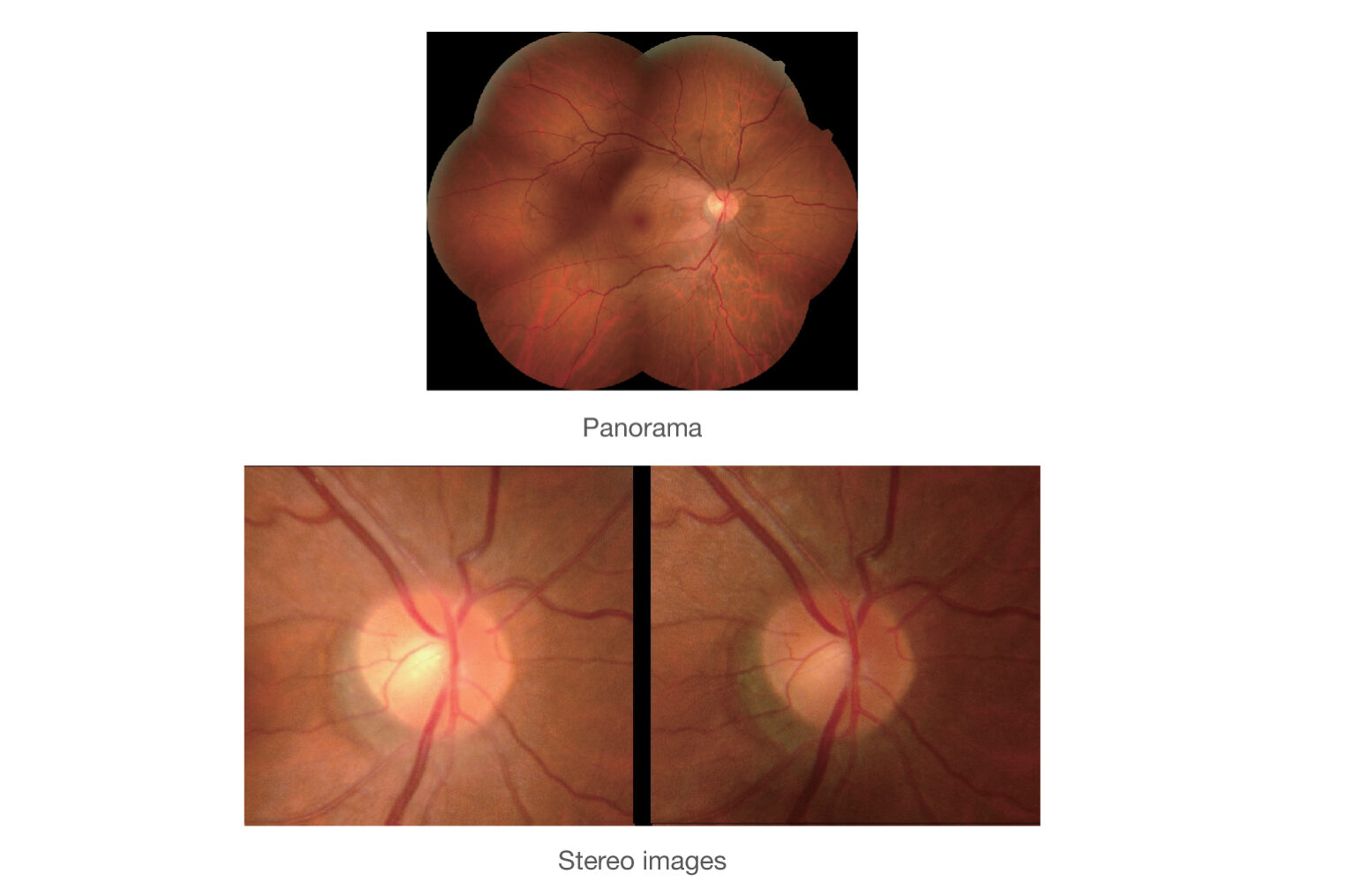
Stereo and panorama photography
The Retina Scan Duo™ 2 includes a built-in 12-megapixel CCD camera, producing high quality fundus images with a 45° angle of view.
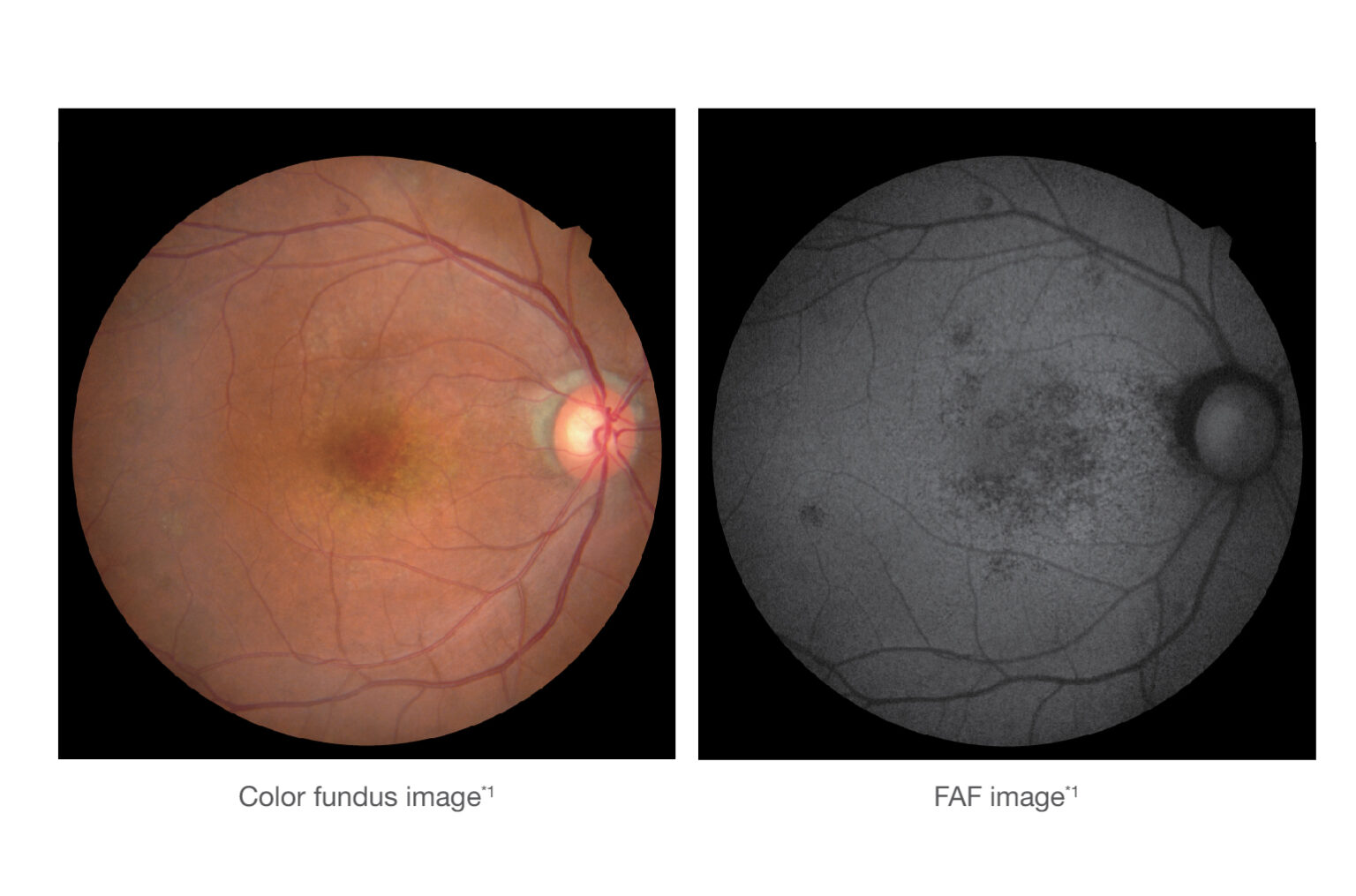
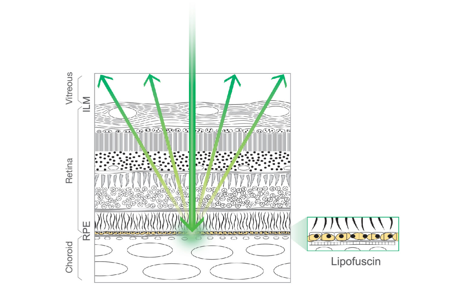
The FAF function is an advanced screening feature that allows non-invasive evaluation of the RPE without contrast dye. FAF is naturally emitted due to the presence of a substance called lipofuscin in the RPE cells. When stimulated with a specific wavelength of light, lipofuscin fluoresces and its distribution can be mapped.
Optional Features
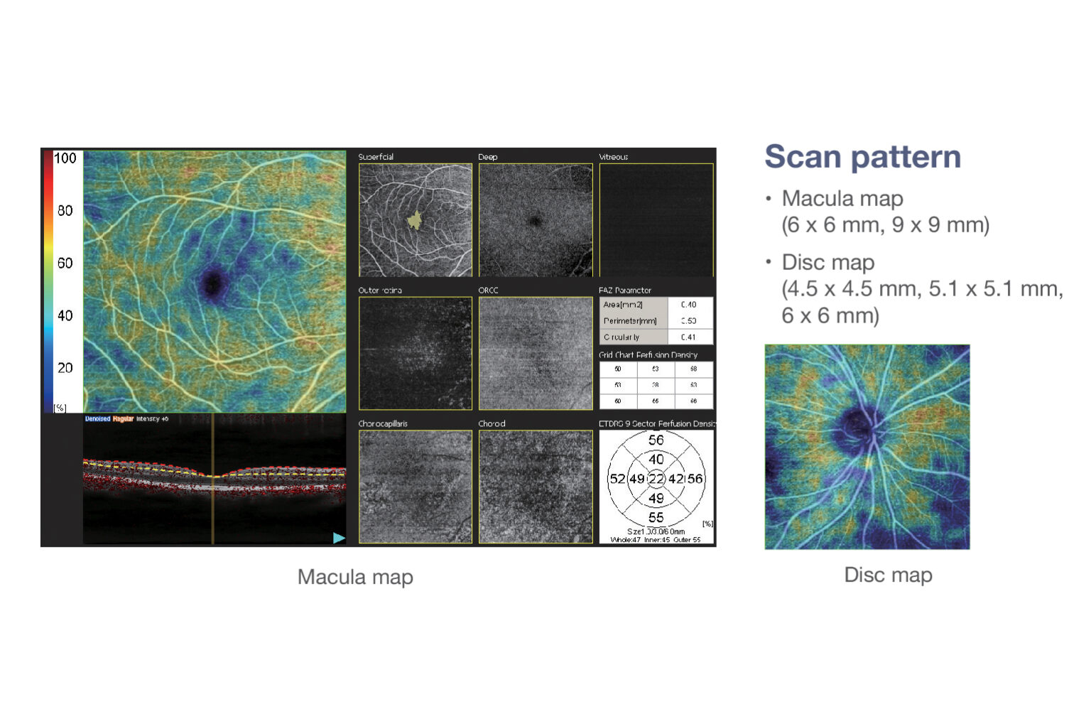
The optional AngioScan is available for OCT-Angiography imaging and diagnostics. The easy to use interface provides seven slabs for the macula map and four slabs for the disc map. This interface has intuitive functionality and removes projection artifacts. Segmentation into multiple slabs allows enhanced assessment of retinal microvasculature at specific depths and regions of interest. The effect of pathology can be evaluated in greater detail at each retinal depth.
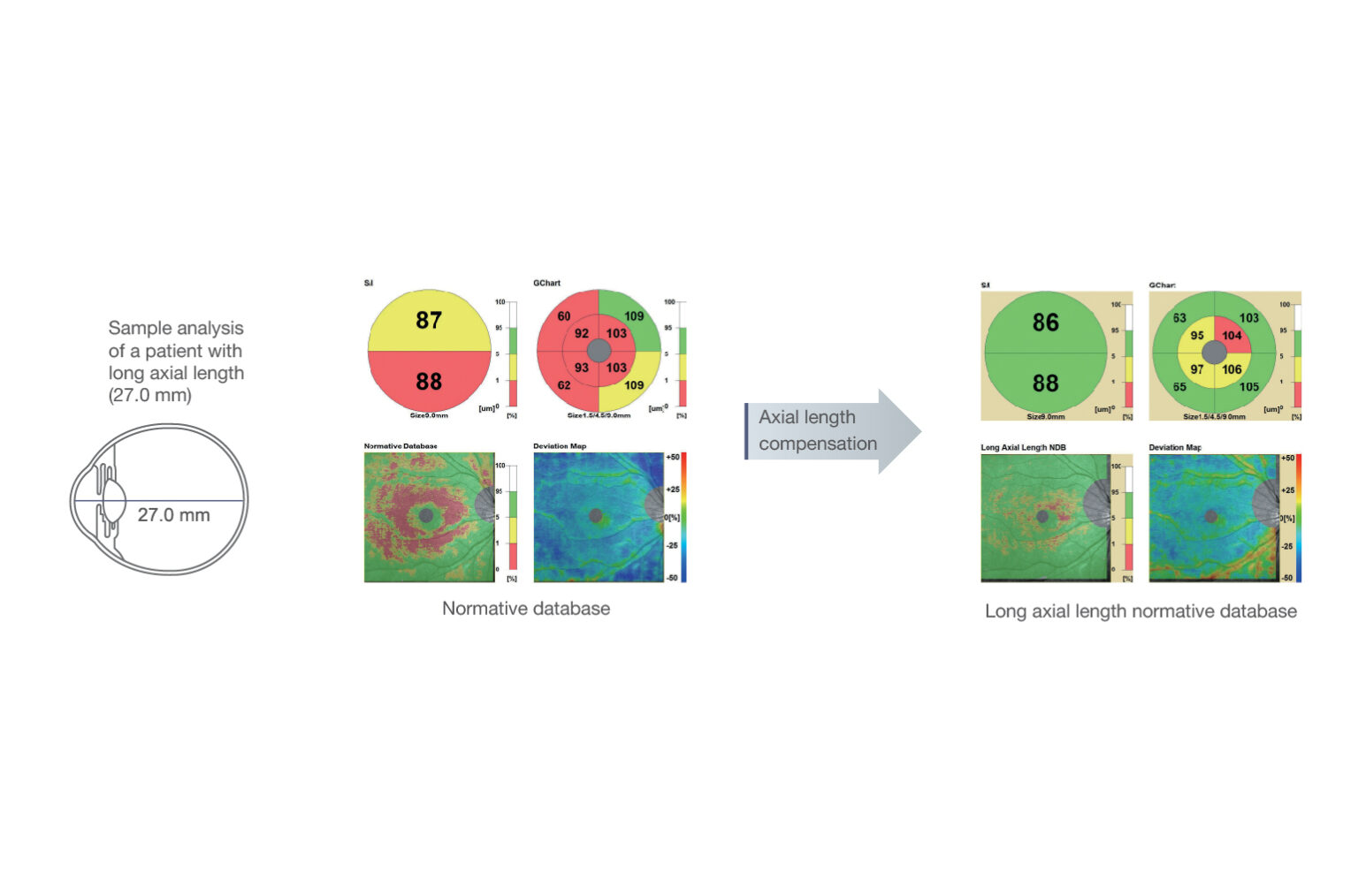
The optional long axial length normative database assists in diagnosing macular diseases and glaucoma in patients with long axial lengths. Data was collected from a sample of Asian patients

The optional anterior segment adapter enables observation and analyses of the anterior segment.
User Friendly Features
The acclaimed 3D auto tracking and auto shot functions allow easy imaging of the fundus. Once alignment is completed, both the OCT and fundus images can be captured in a single shot.
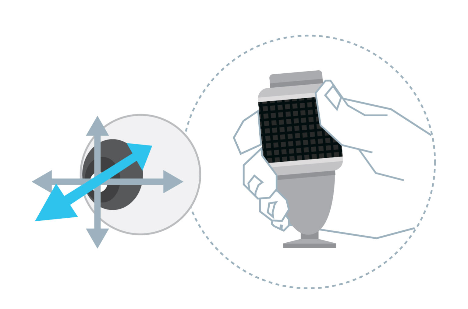
The joystick helps the operator make fine adjustments during alignment and is especially useful in cases of poor fixation that cannot be tracked with automated tracking systems.
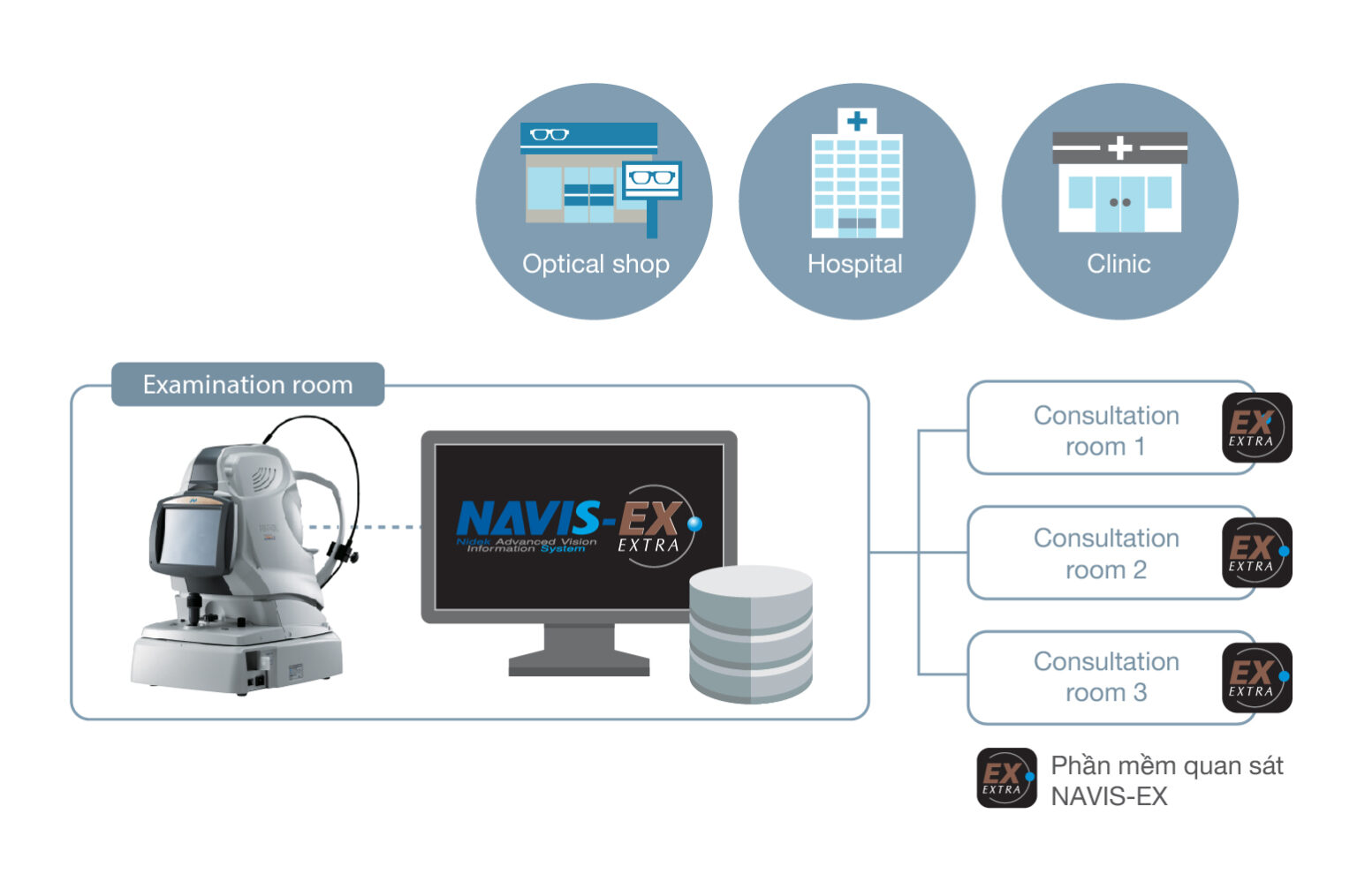
NAVIS-EX is image filing software, which networks the Retina Scan Duo™ 2 and other NIDEK diagnostic devices. This functionality enhances the capability of the diagnostic device with additional features and increases clinical efficiency.
VIETCAN SERVICE & TRADING J.S.C
Address: Room 4A, JVPE Building, Quang Trung Software Park, Tan Chanh Hiep Ward, District 12, Ho Chi Minh City, Vietnam
License No. 0302438819, 11th additional issuance on March 27, 2018.
Place of issue: Department of Planning and Investment HCMC.
Phone: +84 28 6290 8200 – Email: [email protected]

Copyright by VietCan 2019