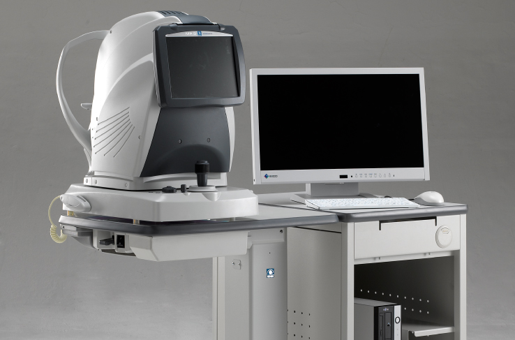
Microperimeter
MP-3
Brand: Nidek – Japan
- The Automatic Microperimeter
- With A Non-Mydriatic
- Fundus Camera
Brand: Nidek – Japan
The MP-3 has a wider range of stimulus intensity, from 0 to 34 dB, compared to the MP-1. The MP-3 measures perimetric threshold values, even for normal eyes. A maximum stimulus luminance of 10,000 asb* allows evaluation of low-sensitivity.
*Complies with the ISO 12866 requirements.
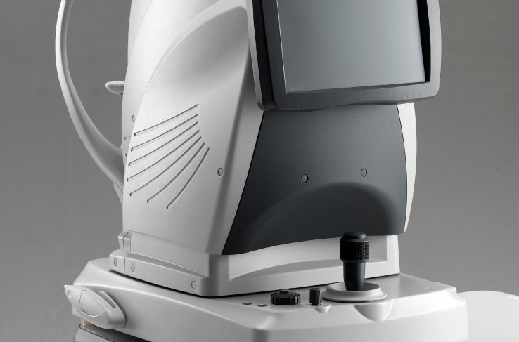
The 12-megapixel fundus camera in the MP-3 acquires high resolution images of retinal pathology and allows easy image acquisition.
Several measurement modes are available for evaluating a variety of pathology including:
A follow-up test can be performed on the same area using the same parameters as a previous test. This feature allows evaluation of disease progression or assessment of pre- and post-treatment outcomes. Any differences in two microperimetry images are displayed for quick, intuitive interpretation.
The MP-3 indicates the percentage of fixation points within 2° and 4° in diameter help confirm fixation stability.
After completion of measurements, results can be evaluated in a specific region of interest to allow easier comparison with other pathology images. By specifying the region of interest, the average results in the region are displayed.
Auto tracking and auto alignment functions provide more accurate measurements increasing patient and operator comfort and efficiency.
These functions allow easy follow-up and reduce variations between examiners, resulting in well-aligned follow up exams.
The MP-3 can measure fixation and determine the preferred retinal locus, simply by having the patient fixate on a target. Any change in fixation can be compared pre- and post-treatment because the patient’s eye is constantly tracked during microperimetry. This test allows evaluation of fixation in patients with central visual field defects and determines whether fixation improved after treatment.
VIETCAN SERVICE & TRADING J.S.C
Address: Room 4A, JVPE Building, Quang Trung Software Park, Tan Chanh Hiep Ward, District 12, Ho Chi Minh City, Vietnam
License No. 0302438819, 11th additional issuance on March 27, 2018.
Place of issue: Department of Planning and Investment HCMC.
Phone: +84 28 6290 8200 – Email: [email protected]

Copyright by VietCan 2019